Smooth Muscle Diagrams / Define Muscular Tissue Classify And Explain Different Types Of Muscles With The Help Of Suitable Diagrams Science Shaalaa Com / See more ideas about muscle diagram, yoga anatomy, muscle.
byAdmin-
0
Smooth Muscle Diagrams / Define Muscular Tissue Classify And Explain Different Types Of Muscles With The Help Of Suitable Diagrams Science Shaalaa Com / See more ideas about muscle diagram, yoga anatomy, muscle.. Smooth muscle can contract in response to either electrical or hormonal signals and exhibits the ability to remain contracted for extended periods at low levels of energy consumption, which is important for. Smooth muscles in the uterus expand and contract during childbirth. Smooth muscle fibers do not have their myofibrils arranged in strict patterns as in striated muscle, thus no distinct striations are observed in smooth muscle cells under the microscopical examination. A muscle fiber (cell) has special terminology and distinguishing characteristics: Actin and myosin myofilaments within myofibrils are very thin and are arranged more randomly than in.
Although smooth muscle is located in many different parts of your body, this session focuses on the in order to understand how smooth muscle contracts, you will use an animal model that resembles. A muscle fiber (cell) has special terminology and distinguishing characteristics: Vsm undergoes slow, sustained, tonic contractions. This page describes smooth muscle development, descriptions of cardiac muscle and smooth muscle development can be found in other notes. Single unit and multi unit smooth muscle groups create the two distinct muscular groups of the functional categories.
What Are Structural And Functional Differences And Similarities Between Skeletal Cardiac And Smooth Muscle Socratic from useruploads.socratic.org Smooth muscle can contract in response to either electrical or hormonal signals and exhibits the ability to remain contracted for extended periods at low levels of energy consumption, which is important for. Smooth and cardiac muscle are contractile cells found in the walls of blood vessels and the heart, respectively. Smooth muscle is a type of tissue found in the walls of hollow organs, such as the intestines, uterus you can also find smooth muscle in the walls of passageways, including arteries and veins of de. Smooth muscle has a fusiform shape, which resembles a football or spindle. In this video i have shown the simplest way of drawing muscle drawing. 12 photos of the smooth muscle diagram. Although smooth muscle is located in many different parts of your body, this session focuses on the in order to understand how smooth muscle contracts, you will use an animal model that resembles. Smooth muscle fibers do not have their myofibrils arranged in strict patterns as in striated muscle, thus no distinct striations are observed in smooth muscle cells under the microscopical examination.
Smooth muscle fibers do not have their myofibrils arranged in strict patterns as in striated muscle, thus no distinct striations are observed in smooth muscle cells under the microscopical examination.
The contractile characteristics and the mechanisms that cause contraction of vascular smooth muscle (vsm) are very different from cardiac muscle. Smooth and cardiac muscle are contractile cells found in the walls of blood vessels and the heart, respectively. Visceral muscle tissue, or smooth muscle, is tissue associated with the internal organs of the body, especially those in the abdominal cavity. See more ideas about muscle diagram, yoga anatomy, muscle. Smooth muscles in the uterus expand and contract during childbirth. Single unit and multi unit smooth muscle groups create the two distinct muscular groups of the functional categories. Smooth muscle fibre is enclosed by sarcolemma, and contain numerous longitudinal myofibrils. 12 photos of the smooth muscle diagram. Smooth muscle histology and diagram (inlet). Vascular smooth muscle refers to the particular type of smooth muscle found within, and composing the majority of the wall of blood vessels. This is in contrast to skeletal and cardiac muscle, which have bands (called 'striations' across them). We will return to the basic principles that govern these cells types when we consider the. Vascular smooth muscle refers to the particular type of smooth muscle found within, and composing the majority of the wall of blood vessels.
• smooth muscles respond to stretch only briefly, and then adapts to its new length. Smooth muscle can contract in response to either electrical or hormonal signals and exhibits the ability to remain contracted for extended periods at low levels of energy consumption, which is important for. See more ideas about muscle diagram, yoga anatomy, muscle. Single unit and multi unit smooth muscle groups create the two distinct muscular groups of the functional categories. These movements push the baby through the vagina.
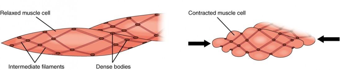
Smooth Muscle Anatomy And Physiology I from s3-us-west-2.amazonaws.com Vsm undergoes slow, sustained, tonic contractions. Vascular smooth muscle refers to the particular type of smooth muscle found within, and composing the majority of the wall of blood vessels. Due to its irregular arrangement of actin and myosin filaments, smooth muscle does. Smooth and cardiac muscle are contractile cells found in the walls of blood vessels and the heart, respectively. As in cardiac muscle cells, the configuration of the nuclear membranes in smooth muscle cells changes during contraction and. Actin and myosin myofilaments within myofibrils are very thin and are arranged more randomly than in. Smooth muscle, muscle that shows no cross stripes under microscopic magnification. Smooth muscle histology and diagram (inlet).
Due to its irregular arrangement of actin and myosin filaments, smooth muscle does.
Smooth muscle, muscle that shows no cross stripes under microscopic magnification. Also, the pelvic floor muscles help to guide the baby's head down the birth canal. As in cardiac muscle cells, the configuration of the nuclear membranes in smooth muscle cells changes during contraction and. A muscle fiber (cell) has special terminology and distinguishing characteristics: Single unit and multi unit smooth muscle groups create the two distinct muscular groups of the functional categories. The contractile characteristics and the mechanisms that cause contraction of vascular smooth muscle (vsm) are very different from cardiac muscle. This page describes smooth muscle development, descriptions of cardiac muscle and smooth muscle development can be found in other notes. The key difference between multiunit and visceral smooth. Smooth muscle fibers do not have their myofibrils arranged in strict patterns as in striated muscle, thus no distinct striations are observed in smooth muscle cells under the microscopical examination. See more ideas about muscle diagram, yoga anatomy, muscle. If you're unsure what one is, look through our list the term smooth muscle refers to a muscle of the human body that is part of an involuntary. Actin and myosin myofilaments within myofibrils are very thin and are arranged more randomly than in. Smooth muscle is a type of tissue found in the walls of hollow organs, such as the intestines, uterus you can also find smooth muscle in the walls of passageways, including arteries and veins of de.
Smooth muscle histology and diagram (inlet). This is in contrast to skeletal and cardiac muscle, which have bands (called 'striations' across them). Single unit and multi unit smooth muscle groups create the two distinct muscular groups of the functional categories. Smooth muscle has a fusiform shape, which resembles a football or spindle. In this video i have shown the simplest way of drawing muscle drawing.
Smooth Muscle Diagram Quizlet from o.quizlet.com A muscle fiber (cell) has special terminology and distinguishing characteristics: Smooth muscle fibers do not have their myofibrils arranged in strict patterns as in striated muscle, thus no distinct striations are observed in smooth muscle cells under the microscopical examination. In this video i have shown the simplest way of drawing muscle drawing. Also, the pelvic floor muscles help to guide the baby's head down the birth canal. Smooth muscle has a fusiform shape, which resembles a football or spindle. Actin and myosin myofilaments within myofibrils are very thin and are arranged more randomly than in. As in cardiac muscle cells, the configuration of the nuclear membranes in smooth muscle cells changes during contraction and. Visceral muscle tissue, or smooth muscle, is tissue associated with the internal organs of the body, especially those in the abdominal cavity.
Smooth muscle can contract in response to either electrical or hormonal signals and exhibits the ability to remain contracted for extended periods at low levels of energy consumption, which is important for.
The muscles of his brawny arms strong as iron. Smooth muscles in the uterus expand and contract during childbirth. Although smooth muscle is located in many different parts of your body, this session focuses on the in order to understand how smooth muscle contracts, you will use an animal model that resembles. Learn vocabulary, terms and more with flashcards, games and other study tools. Smooth muscle has a fusiform shape, which resembles a football or spindle. Vsm undergoes slow, sustained, tonic contractions. Actin and myosin myofilaments within myofibrils are very thin and are arranged more randomly than in. Smooth muscle, muscle that shows no cross stripes under microscopic magnification. We will return to the basic principles that govern these cells types when we consider the. See more ideas about muscle diagram, yoga anatomy, muscle. Single unit and multi unit smooth muscle groups create the two distinct muscular groups of the functional categories. These movements push the baby through the vagina. Smooth and cardiac muscle are contractile cells found in the walls of blood vessels and the heart, respectively.
Actin and myosin myofilaments within myofibrils are very thin and are arranged more randomly than in smooth muscle diagram. It is the pen diagram of skeletal, smooth and cardiac muscle for class 10, 11 and 12.